How to Do a Blood Draw on a Llama
Llama Venipuncture - Drawing Blood in the Llama
To view these resources with no ads, please Login or Subscribe (and help support our site).
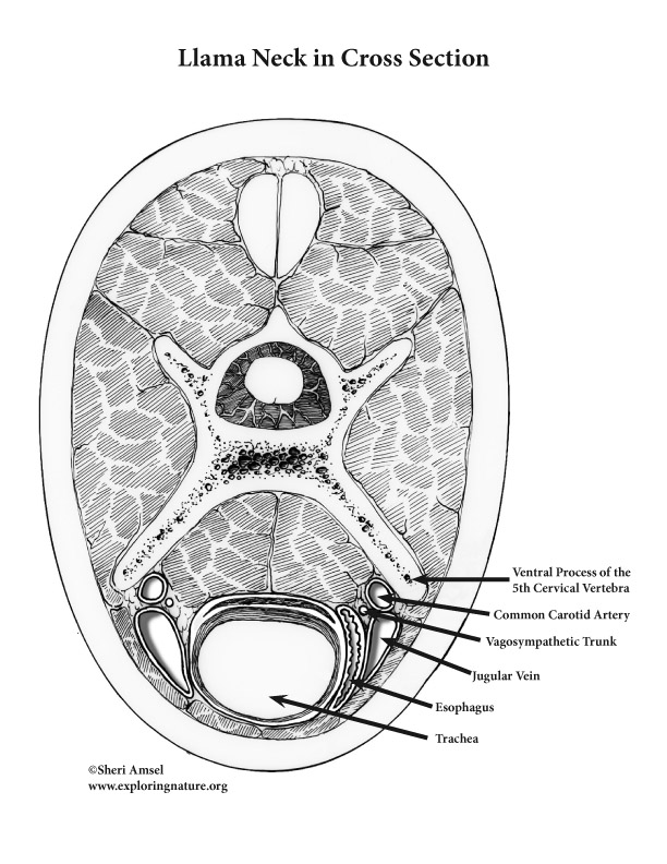
Drawing blood ( venipuncture ) in the llama is more difficult than in common domestic animals. Their wooly fleece impairs visibility when you try to locate the jugular vein. For aesthetic reasons, llamas are not routinely clipped to imporve visibility. The optimal site for venipuncture will be described with the aid of several illustrations depicting the anatomy of the llama's blood vessels and other structures in their long neck (anatomy of the cervical region). We hope this is a helpful guide.
The positions of the jugular vein, common carotid artery, vagosympathetic trunk, and esophagus in association with the unique cervical vertebra in the llama (longer than most mammals), necessitates careful examination to locate a site for venipuncture.

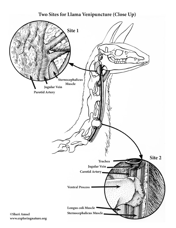
Two potential sites for drawing blood are suggested.
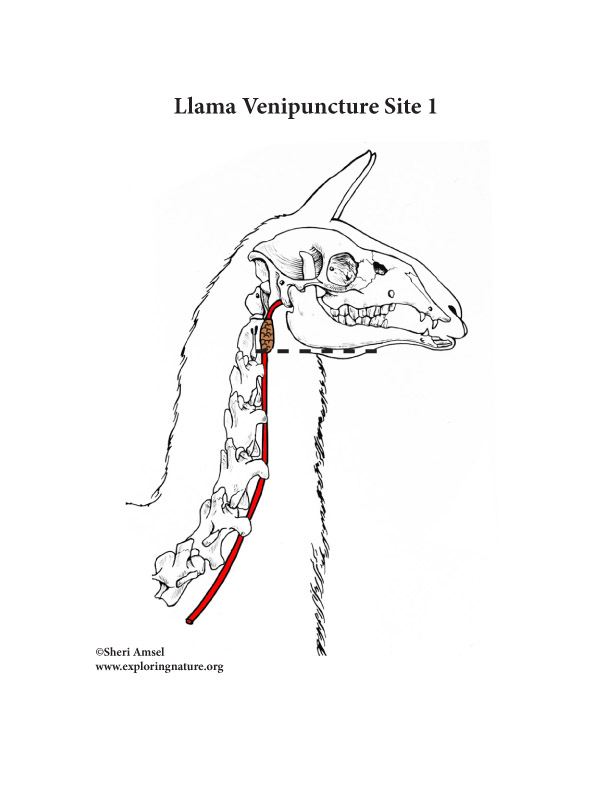
Site one is in the cranial portion of the neck at the level of the mandible. A line continuous with the ventral border of the mandible is extended 3-4 cm dorsad from the angle of the jaw. At this site, an 18 gauge, 1.5 inch needle can be thrust through the cervical skin, platysma muscle and variably, the parotid salivary gland into the jugular artery.
The advantage to venipuncture in this region of the neck is that the jugular vein is superficial to the sternocephalicus muscle and is not as close to the carotid artery as in the caudal part of the neck. The disadvantage in this location is that here the llama's ski can be up to one centimeter thick. At this level, the needle may also pass through the parotid salivary gland (see illustration). lastly, there is a real possibility of head movement if the animal is not securely restrained.

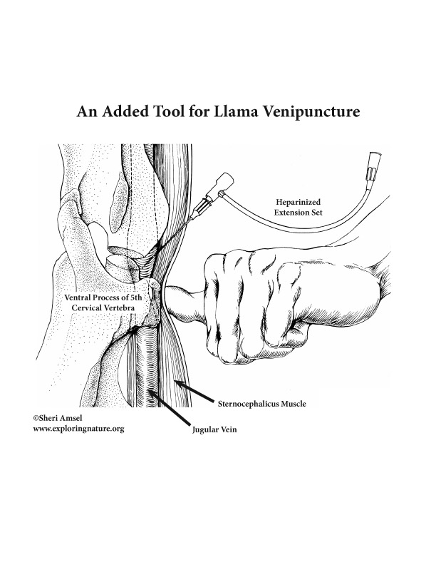
The second site is in the caudoventral portion of the neck where the ventral process of the cervical vertebrae can be palpated. the processes of the 5th and 6th cervical vertebra are most prominent. Palpating craniad from the point of the shoulder, the 2nd prominence felt will be the ventral process of the 5th cervical vertebra. This process should be palpated with the thumb and then the thumb can be slid into a depression just medical to the process superficial to the jugular vein, where it courses between the sternocephalicus muscle and the ventral process of the vertebra.
Vigorously stroke over the vein, cranial to the site of compression, to assist in its identification, which is assured by transmission of pressure waves to yout compressing thumb. Venipuncture should be accomplished readily immediately cranial to the site of compression using an 18 gauge, 1.5 inch needle directly craniad at the angle of 45° or less to the plane of the cervical skin.
The advantages at this site are that the skin is on 2-5 mm thick and the neck is relatively stable when the animal moves its head. The jugular vein can be compressed and may be detected enlarging cranial to the compression site for positive identification. The disadvantage here is that this position necessitates the operator's body to be lower and in potential jeopardy without adequate restraint. Also, the jugular vein and common carotid artery are in proximity and the artery could be punctured.
An added confusion at either site is that venous blood of the llama bears a high percentage of oxyhemoglobin, so is very red. Arterial blood, however, is unmistakable, due to the pulsatile flow when the common carotid artery is punctured. On occasion, the jugular valves will interfere with aspiration of the blood.


A technique advantageous at both sites uses a heparinized extension set connected to the needle. This allows blood to flow into the tubing and examined before attaching the syringe. Because of the 8-12 cm of tubing on an extension set, the llama can move without dislodging the needle.

Please Login or Subscribe to access downloadable content.
To view these resources with no ads, please Login or Subscribe (and help support our site).
Acknowledgements:
Written and illustrated by Sheri Amsel, MS (Veterinary Anatomy and Medical Illustration) as published in Veterinary Medicine Journal in 1987.
Assistance in research, dissection and anatomy from Dr. Robert Kainer (CSU Veterinary School), Dr. LaRue Johnson (CSU Veterinary School) and Dr. Murray Fowler (UC Davis).
Citing Research References
When you research information you must cite the reference. Citing for websites is different from citing from books, magazines and periodicals. The style of citing shown here is from the MLA Style Citations (Modern Language Association).
When citing a WEBSITE the general format is as follows.
Author Last Name, First Name(s). "Title: Subtitle of Part of Web Page, if appropriate." Title: Subtitle: Section of Page if appropriate. Sponsoring/Publishing Agency, If Given. Additional significant descriptive information. Date of Electronic Publication or other Date, such as Last Updated. Day Month Year of access < URL >.
Here is an example of citing this page:
Amsel, Sheri. "Llama Venipuncture - Drawing Blood in the Llama" Exploring Nature Educational Resource ©2005-2022. January 4, 2022
< http://www.exploringnature.org/db/view/Llama-Venipuncture-Drawing-Blood-in-the-Llama >
How to Do a Blood Draw on a Llama
Source: https://www.exploringnature.org/db/view/Llama-Venipuncture-Drawing-Blood-in-the-Llama
0 Response to "How to Do a Blood Draw on a Llama"
Enviar um comentário